 Evolution
 Intelligent Design
The Mystery of Vision
Editor’s note: Physicians have a special place among the thinkers who have elaborated the argument for intelligent design. Perhaps that’s because, more than evolutionary biologists, they are familiar with the challenges of maintaining a functioning complex system, the human body. With that in mind, Evolution News is delighted to offer this series, “The Designed Body.” For the complete series, see here. Dr. Glicksman practices palliative medicine for a hospice organization.
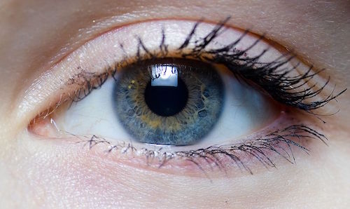
 Everyone knows that an odometer measures distance and a speedometer measures velocity. But how do they do it? Each device is essentially a sensory transducer with a mechanism that enables it to sense a physical phenomenon and convert it into useful information. The body has sensory transducers as well that it uses to detect physical phenomena and know what is going on within and without. Vision is the sensation we experience when light, usually reflecting off an object that is within a very narrow range of frequency, enters our eyes.
Common sense teaches that without this special sense our earliest ancestors could never have survived. Evolutionary biologists claim that the presence of different light-sensitive organs in early life forms made it easy for chance and the laws of nature alone to bring about vision. But just like the development of various inventions and technologies, all human experience teaches that intelligent design is a much more plausible explanation. The position of Darwinists not only oversimplifies the development of the irreducibly complex eye, but also does not take into account how our brain converts what it receives from our eyes so that we experience vision.
Nobody, not even evolutionary biologists, truly understands this mystery. The fact that nobody understands it should make any scientist wary of claiming to know how the eye and vision came into being. Yet Darwinists rush in to do just that. Let’s look at what makes up the eye, how it works, what the brain receives from it, and how it converts that information into the sensation we call sight.
The human eye is a very complex sensory organ in which many parts work together to focus light on its retina. Although it is in the retina where the nerve impulses for vision begin, the other parts of the eye play important roles that support and protect retinal function. The five different bones that make up the orbital cavity protect about two-thirds of the eyeball and provide the base for the origin tendons of the muscles responsible for eye movement. The eyelids and lashes protect the eye from exposure to too much light or dust, dirt, bacteria, and other foreign objects. A film of tears, consisting of oil, water, and mucus is produced by the oil glands of the eyelids, the lacrimal gland, and the conjunctiva that overlies the sclera (the white outer protective coating of the eyeball). The tear film lubricates the eye, protects it from infection and injury, nourishes the surrounding tissue, and preserves a smooth surface to aid in light transmission.
The cornea is a transparent connective tissue that protects the front of the eye while allowing light to enter. The cornea is transparent because it lacks blood vessels (avascular), instead receiving oxygen, water, and nutrients from two sources. One is the tears that constantly wash across it by the blinking eyelids, and the other is the clear fluid (aqueous humor) within the anterior chamber that sits behind the cornea and in front of the lens. Light rays that reflect from an object more than twenty feet away enter parallel to each other and must be bent (refracted) to focus them on the area in the retina for central (macula) and sharp vision (fovea). The cornea’s curvature plays a major role in focusing the light that enters the eye onto the retina.
The lens is a transparent, elastic biconvex structure that is kept in place by suspensory ligaments. Like the cornea, it is avascular and obtains its oxygen, water, and nutrients from the aqueous humor in the anterior chamber. As noted above, light rays from a distance (greater than twenty feet) enter the eye in parallel, whereas those from nearby (generally less than twenty feet away) spread out. To focus the light on the macula and fovea, this diverging light must be further refracted and the biconvex curvature of the lens accomplishes this task. Since what the eye focuses on close-up is always changing, the curvature of the lens can be reflexively adjusted (accommodation) so that the light rays will strike the retina in the area for sharp vision.
The choroid is the layer of tissue located between the sclera and the retina and provides the circulation to the back of the eye. The choroid also contains the retinal pigmented epithelium, which sits behind the retina and absorbs light. This prevents light from reflecting back on the photoreceptors and causing visual blurring. The extension of the choroid in the front of the eye is the colored iris, consisting of two different muscles that control the amount of light that enters through its opening (pupil).
Finally, the thick, transparent, and gelatinous substance that forms and shapes the eyeball is the vitreous. It is able to compress and return to its natural position, allowing the eyeball to withstand most physical stresses without serious injury.
Each eye has about one hundred twenty million rods arranged throughout the retina. The rods contain a photopigment called rhodopsin which is very sensitive to all the wavelengths of the visible light spectrum. In contrast, there are only about six million cones that are mostly concentrated in the macula, primarily in the cone-only fovea. Each cone contains one of three different photosensitive pigments, called photopsins, which tend to react stronger to either the red, green, or blue wavelengths of light. Both rhodopsin and the photopsins are dependent on Vitamin A.
When photons of light strike the retina they interact with the photoreceptor cells and cause an electrical change and the release of a neurotransmitter. Messages are passed through interconnecting neurons within the retina. These retinal interneurons process the information and send the resulting nerve signals along the optic nerve to the brain. About eighty percent of the optic nerve impulses travel to neurons within the brain. These pass on the sensory information to the visual cortex in the occipital lobes. However, the remaining twenty percent veer off and provide sensory data to the neurons in the brainstem that service muscles that help the eye to function better and provide protection.
For example, if you enter a dark room, the dilating muscle of the iris immediately contracts, causing the pupil to enlarge. This lets more light into the eye to help improve vision. But if you shine a bright light into your eye, the contracting muscle of the iris instantly goes into action, causing the pupil to diminish in size to protect the retina from too much light. This is called the pupillary light reflex, which is often used by physicians to determine the presence of brainstem function.
In considering the nature of the sensory data being presented from the eyes to the visual cortex, several points must be kept in mind. First, the use of the cornea and lens to refract and focus light on the retina results in a reversed and upside-down image. This means that what appears in the right upper half of the visual field is detected by the left lower half of the retina and what appears in the left lower half of the visual field is detected by the right upper half of the retina etc. Second, looking through one eye shows that there is an overlap in the nasal visual fields (the right half of the left eye and the left half of the right eye). This overlap provides the visual cortex with two different perspectives and allows for depth perception.
Finally, impulses sent along each optic nerve split-up on their way to the brain. The messages from the nasal half of the retina cross over from right to left and from left to right through what is called the optic chiasm. However the impulses from the temporal half of the retina (the left half of the left eye and the right half of the right eye) stay on the same side. This means that everything seen by the right half of each eye (the nasal field of the left eye and the temporal field of the right eye) goes to the left occipital lobe and everything seen by the left half of each eye (the nasal field of the right eye and the temporal field of the left eye) goes to the right occipital lobe. Our brain then takes this upside down, turned around, split-up and overlapping collection of photon-generated nerve impulses and provides us with what we experience as vision. How it is able to accomplish this feat remains entirely unknown.
If you have ever used a magnifying glass to focus light onto a paper to make it burn, then you know that the refractive power of a lens is dependent on its degree of curvature, which is inversely related to the distance it takes to bring the light together at a focal point. The higher the refractive power, the shorter the focal distance, and vice versa. The eye is dependent on the combined refractive power of the cornea and the lens (58 diopters) to focus light onto the area of the retina for sharp vision. And as luck would have it, the distance from the cornea to the retina (23 mm) is exactly what it should be to get the job done. What do you know?
For our earliest ancestors to have been able to safely find food and water and properly prepare and handle it for ingestion, would have required them having normal distance and near vision. Eye doctors know that about a four percent increase in the combined refractive power of the cornea and lens (or a lengthening of the eye) results in severe myopia (not being able to see the big E on the eye chart clearly). And a twenty five percent decrease in both of these leads to difficulties with distance and near vision.
When evolutionary biologists talk about vision, not only do they leave out how it is irreducibly complex (all of the parts of the eye and the brain are needed for proper function) but also that it demonstrates natural survival capacity, in that the combined refractive power of the cornea and lens and the lens’s ability to adjust to close-up objects perfectly matches the diameter of the eyeball. Remember, when it comes to life and the laws of nature, real numbers have real consequences. Without the right refractive power or eyeball diameter our earliest ancestors would have been as blind as bats.
But in that case, as some people mistakenly argue, evolution would have just made them develop sonar instead, because that would have been what they needed to survive. Next time we’ll look at hearing.
Photo credit: Laitr Keiows (Own work) [CC BY-SA 3.0 or GFDL], via Wikimedia Commons.