 Evolution
 Intelligent Design
 Medicine
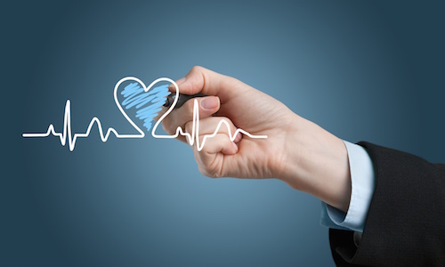
The Cardiovascular System: Regulating Heart Rate
Editor’s note: Physicians have a special place among the thinkers who have elaborated the argument for intelligent design. Perhaps that’s because, more than evolutionary biologists, they are familiar with the challenges of maintaining a functioning complex system, the human body. With that in mind, Evolution News & Views is delighted to present this series, “The Designed Body.” For the complete series, see here. Dr. Glicksman practices palliative medicine for a hospice organization.
The job of the heart is to pump blood throughout the body to meet its metabolic needs. However, since blood has mass, this means the heart must have enough power to move it against natural forces like inertia, friction, and gravity.
Cardiac output is directly related to how much blood the heart pumps out with each contraction (stroke volume) and how fast the heart beats per minute (heart rate). At rest, with a stroke volume of 70 mL and a heart rate of 72 bpm, the cardiac output is about five liters per minute (70 x 72). However, with increased muscle activity and physical effort, the autonomic nervous system stimulates the heart to pump harder and faster so the cardiac output can rise to the occasion.
Clinical experience tells us that our earliest ancestors would have needed a cardiac output of at least 25 liters per minute in order to carry out activities needed to survive and reproduce. The normal heart, under the influence of the sympathetic nerves, accomplishes this by increasing its stroke volume to 125 mL and its heart rate to 200 bpm (125 x 200). The last few articles in this series have shown that to accomplish the task, the heart must have enough blood flow to its myocardium. Its valves must let blood flow freely forward without letting any go backward, and the ventricles must contract and relax properly.
Evolutionary biologists would have us believe that all of this came about by chance and the laws of nature alone. However, clinical experience teaches that real numbers have real consequences. Without wide enough coronary arteries for adequate blood flow to the heart muscle, open enough valves for forward blood flow, tight enough closed valves to prevent backward blood flow, and adequate ventricular contraction and relaxation for a large enough stroke volume, the cardiac output needed to meet the metabolic needs of our earliest ancestors could never have been achieved.
But there is one more very important component of heart function. Here I will review how real numbers affect the heart’s electrical system.
As you may recall, since K+ ions tend to leak out of the cell through their specific ion channels more than Na+ ions do through theirs, the inside of the plasma membrane has as a negative electrical charge and the outside has a positive one. This difference in the electrical charge between the inside and the outside of the cell is called the resting membrane potential.
Nerve and muscle cells are considered excitable because, when properly stimulated, they are able to reverse this situation, by making the inside positive and the outside negative, in a process called depolarization. After this happens, they undergo repolarization to return to the resting membrane potential. In general, skeletal muscle requires stimulation by a nerve to cause depolarization and muscle contraction.
However, although nerves can modulate how often and how hard the heart contracts, there are cells within the heart that can depolarize on their own without the need for direct nerve stimulation. In particular, the right atrium houses the sinus node (sino-atrial or SA node) which depolarizes faster than all of the others. This makes it the natural pacemaker of the heart and, at rest, generally makes the heart contract at 60-100 beats per minute (bpm). The electrical message from the sinus node stimulates the atria to contract and moves quickly to a junction box that sits between the atria and the ventricles called the atrio-ventricular node (AV node).
The AV node is the electrical wiring that physically connects the atria to the ventricles. Like the sinus node, the AV nodal tissue can depolarize automatically and generate its own electrical impulse. But its intrinsic rate is only 40-60 bpm. Since the sinus node intrinsically has a faster depolarization rate than the AV node, its electrical impulse depolarizes the AV node before it can generate its own impulse. The AV node slows the electrical impulse by about 1/10 of a second, allowing the atria to contract fully, and then sends the message to specialized conducting tissue. This tissue then rapidly conducts the electrical impulse to the ventricles so that coordinated contraction and the pumping of blood out of the heart can take place.
Like the sinus and AV nodes, the specialized conducting tissue below the AV node can also automatically depolarize. However, its intrinsic rate is only 20-40 bpm and so it is normally stimulated from above before it can generate its own impulse. It is important to note that this hierarchical set up for impulse formation and conduction is what allows for coordinated ventricular contraction and normal cardiac function. Let’s see what can happen when the electrical system isn’t working right.
Normal sinus rhythm is when the sinus node paces the heart at 60-100 bpm and sinus bradycardia is when it paces it at less than 60 bpm. It is not unusual for a person to have a heart rate of 45-50 bpm at complete rest and even 30-40 bpm during sleep. However, if the heart rate stays below 45 bpm while awake, and in particular, if it does not increase adequately during physical activity, then something is wrong.
This is called a bradyarrhythmia and in the absence of medication or hypothyroidism, is usually due to either a defect in sinus node function (sick sinus syndrome)or a problem with conduction (atrioventricular heart block). The commonest causes for these conditions are age-related degeneration and coronary artery disease.
The normal cardiac output (CO) at rest is about five liters per minute and is directly related to the heart rate (HR), which is usually about 72 bpm. This means that a bradyarrhythmia of 35 bpm, at rest, would cut the CO in half to two and a half liters per minute. It is possible to live with such a slow heart rate and low cardiac output, as long as you do nothing at all. Most people with significant bradyarrythmias are not able to get their heart rate up fast enough, or their cardiac output high enough, to be physically active.
In fact, most people with bradyarrhythmias have problems just walking slowly or going up stairs, in addition to normal, everyday activities. This means that having a bradyarrhythmia would have made it impossible for our earliest ancestors to generate the CO of 25 liters per minute they would have needed to survive and reproduce. So real numbers can mean debility.
A sinus tachycardia is when the sinus node paces the heart at a rate greater than 100 bpm. An increase in metabolic needs, resulting in stimulation of the sympathetic nervous system, is the usual cause. Physical exertion, stress, anxiety, fear, pain, fever, heart failure, hyperthyroidism and low blood pressure are some of the usual culprits. However, conditions such as poor coronary blood flow, emphysema, hypertension and low serum potassium, can predispose some of the ion channels in the heart tissue to malfunction.
These changes can result in abnormalities of depolarization and repolarization which lead to increased electrical irritability of the atria and the ventricles. When this happens, they can take over the pacing of the heart from the sinus node with unusually rapid heart rates that exceed 100 bpm and are called tachyarrhythmias.
The atrial tachyarrhythmias can cause the heart rate to rise to over 200 bpm, significantly reducing the time for relaxation and filling of the ventricles. Since these fast heart rates occur spontaneously at rest and without exercise-induced sympathetic stimulation, the stroke volume and cardiac output can often drop dramatically. People with atrial tachyarrhythmias usually feel a fluttering in the chest along with weakness, dizziness, chest discomfort, and shortness of breath on limited exertion. If our earliest ancestors had had atrial tachyarrhythmias, they never could have survived.
In summary, four different aspects of cardiac function would need to have been working properly to allow for the survival of our earliest ancestors: coronary blood flow, valve function, ventricular contraction and relaxation, and the electrical system. When it comes to survival, life has to follow the rules by taking control and making sure that all the numbers regarding its various functional parameters add up properly.
Evolutionary theory claims to explain how life came into being by describing only how life looks but not how it actually works within the laws of nature. That is a significant drawback that seriously undermines the theory’s value as an explanation of biological origins.
Next, we’ll consider what actually causes the heart to stop beating in cardiac arrest.
Image: � BillionPhotos.com / Dollar Photo Club.